ClearStepsCLEARSTEPS AI
Teacher Login →Student Login →

Human Reproduction: Class 12 Biology Practice Questions

24 original exam-pattern questions with full answers, matched to the current CBSE Class 12 paper design, including case-based questions. Attempt each question before opening the answer — or start a free 14-day trial ↓ for the full bank.

Q1Case-based4 marks

Dr. Meera, a gynaecologist, is counselling a couple who have been trying to conceive for two years without success. During investigation, it was found that the female partner has a complete blockage of both fallopian tubes due to a previous pelvic infection. Her ovaries are fully functional and produce healthy oocytes each month. The male partner has normal semen parameters. Dr. Meera explains the structure and function of the female reproductive tract, particularly the role of the fallopian tube in natural fertilisation, and recommends an appropriate Assisted Reproductive Technology (ART).

Read the following passage and answer the questions that follow.

Dr. Meera, a gynaecologist, is counselling a couple who have been trying to conceive for two years without success. During investigation, it was found that the female partner has a complete blockage of both fallopian tubes due to a previous pelvic infection. Her ovaries are fully functional and produce healthy oocytes each month. The male partner has normal semen parameters. Dr. Meera explains the structure and function of the female reproductive tract, particularly the role of the fallopian tube in natural fertilisation, and recommends an appropriate Assisted Reproductive Technology (ART).

(i) Name the specific region of the fallopian tube where fertilisation normally occurs. What structural feature of the fallopian tube facilitates the movement of the fertilised egg towards the uterus? (1+1=2)

(ii) Since both fallopian tubes are blocked, Dr. Meera recommends a specific ART procedure. Name the ART technique she would most appropriately recommend for this couple, and justify your choice over GIFT (Gamete Intra-Fallopian Transfer). (1+1=2)

Show answer
MARKING SCHEME — Case Study (4 marks)

(i) [2 marks — 1+1]

Value Point 1 (1 mark):
Fertilisation normally occurs in the ampullary region of the fallopian tube (oviduct).
[Also accept: ampulla / ampullary-isthmic junction]

Value Point 2 (1 mark):
The fallopian tube is lined with ciliated epithelium; the beating of cilia (along with peristaltic muscular contractions of the tube wall) propels / moves the fertilised egg (zygote) towards the uterus.
[Accept either cilia OR peristalsis for 1 mark; both together also acceptable for 1 mark]

(ii) [2 marks — 1+1]

Value Point 1 (1 mark):
Dr. Meera would recommend IVF-ET (In Vitro Fertilisation followed by Embryo Transfer) / Test Tube Baby technique.
[Also accept: ZIFT — Zygote Intra-Fallopian Transfer — as a second acceptable ART if the student justifies it differently; however, IVF-ET with uterine transfer is the most appropriate answer given bilateral tubal blockage]

Value Point 2 (1 mark — Justification):
GIFT (Gamete Intra-Fallopian Transfer) requires at least one functional / patent fallopian tube, as gametes are transferred into the tube for fertilisation to occur naturally inside the tube.
Since BOTH fallopian tubes of this patient are completely blocked, GIFT is not possible / feasible.
In IVF-ET, fertilisation is performed outside the body (in vitro) and the resulting embryo is transferred directly into the uterus (ET = Embryo Transfer), bypassing the need for functional fallopian tubes entirely.
[Award 1 mark for a clear, correct biological justification that identifies the requirement of a patent tube for GIFT and/or the bypass mechanism of IVF-ET]
Q2Case-based4 marks

Dr. Meera, a gynaecologist, was counselling a couple undergoing infertility treatment. She explained that after ovulation, the secondary oocyte is released from the Graafian follicle and is swept into the fallopian tube. Fertilisation, if it occurs, takes place in the ampullary region. The fertilised egg then undergoes a series of mitotic divisions as it travels toward the uterus. By the time it reaches the uterus, it has transformed into a hollow, fluid-filled structure that embeds itself into the endometrium. Dr. Meera showed the couple a diagram of the female reproductive tract with four structures labelled P, Q, R, and S at different stages along this journey.

Read the following passage and answer the questions that follow:

Dr. Meera, a gynaecologist, was counselling a couple undergoing infertility treatment. She explained that after ovulation, the secondary oocyte is released from the Graafian follicle and is swept into the fallopian tube. Fertilisation, if it occurs, takes place in the ampullary region. The fertilised egg then undergoes a series of mitotic divisions as it travels toward the uterus. By the time it reaches the uterus, it has transformed into a hollow, fluid-filled structure that embeds itself into the endometrium. Dr. Meera showed the couple a diagram of the female reproductive tract with four structures labelled P, Q, R, and S at different stages along this journey.

(a) Name the process by which the fertilised egg undergoes repeated mitotic divisions without increase in cell size, and name the structure formed when 8–16 cells are produced by this process. (1 mark)

(b) Identify the hollow, fluid-filled structure that implants into the endometrium. Name the two distinct cell layers/regions that make up this structure and state the role of each. (2 marks)

(c) The couple asked Dr. Meera: 'If fertilisation does not occur, what happens to the secondary oocyte and the corpus luteum?' Give a scientifically accurate answer. (1 mark)

Show answer
MARKING SCHEME — Human Reproduction (Case Study) [4 marks]

──────────────────────────────────────────
(a) [1 mark]

• The process is called CLEAVAGE (mitotic divisions of the zygote without growth in cell size). [½ mark]
• The structure formed when 8–16 cells are produced is called MORULA. [½ mark]

(Award full 1 mark for both correct terms; accept 'cleavage / segmentation' for the process.)

──────────────────────────────────────────
(b) [2 marks]

The hollow, fluid-filled structure that implants into the endometrium is the BLASTOCYST. [½ mark]

Two distinct regions of the blastocyst:

┌──────────────────────┬────────────────────────────────────────────────────┐
│ Region / Cell layer │ Role │
├──────────────────────┼────────────────────────────────────────────────────┤
│ Trophoblast │ Outer layer of cells; helps in implantation into │
│ │ the endometrium; later forms the placenta. │
├──────────────────────┼────────────────────────────────────────────────────┤
│ Inner Cell Mass │ Cluster of cells (embryoblast) at one pole; │
│ (Embryoblast) │ gives rise to the embryo proper (all tissues and │
│ │ organs of the foetus). │
└──────────────────────┴────────────────────────────────────────────────────┘

[1 mark for naming both regions correctly + 1 mark for stating the role of each]
(Accept 'outer layer — trophoblast' and 'inner cell mass / embryoblast' with roles as above.)

──────────────────────────────────────────
(c) [1 mark]

If fertilisation does NOT occur:
• The secondary oocyte degenerates and is not fertilised. [½ mark]
• The corpus luteum (formed from the ruptured Graafian follicle after ovulation) degenerates into the corpus albicans (a white fibrous scar tissue); progesterone and oestrogen levels fall, leading to menstruation (shedding of the endometrium). [½ mark]

(Award full 1 mark for both points; accept 'corpus luteum degenerates / breaks down → corpus albicans → progesterone falls → menstruation' as a linked sequence.)

──────────────────────────────────────────
SUMMARY OF MARK ALLOCATION:
(a) Cleavage + Morula — 1 mark
(b) Blastocyst named + Trophoblast (role) + Inner Cell Mass (role) — 2 marks
(c) Degeneration of secondary oocyte + Corpus luteum → Corpus albicans / menstruation — 1 mark
TOTAL: 4 marks
Q3Case-based4 marks

Dr. Priya, a gynaecologist, is consulting two patients on the same day.

Patient A (Mrs. Sharma, 28 years old): She has been trying to conceive for two years. Investigations reveal that her fallopian tubes are completely blocked bilaterally due to a previous infection. Her husband's semen analysis is normal. Her uterus and endometrium are healthy. Her menstrual cycle is regular (28-day cycle).

Patient B (Mrs. Kapoor, 32 years old): She conceived naturally but is now 8 weeks pregnant. Her blood test shows rising levels of a hormone that is keeping her corpus luteum functional, preventing menstruation. She is experiencing morning sickness. An ultrasound shows a developing embryo with a disc-like structure connecting it to the uterine wall, through which nutrients and oxygen are being supplied.

Read the following case carefully and answer the questions that follow:

Dr. Priya, a gynaecologist, is consulting two patients on the same day.

Patient A (Mrs. Sharma, 28 years old): She has been trying to conceive for two years. Investigations reveal that her fallopian tubes are completely blocked bilaterally due to a previous infection. Her husband's semen analysis is normal. Her uterus and endometrium are healthy. Her menstrual cycle is regular (28-day cycle).

Patient B (Mrs. Kapoor, 32 years old): She conceived naturally but is now 8 weeks pregnant. Her blood test shows rising levels of a hormone that is keeping her corpus luteum functional, preventing menstruation. She is experiencing morning sickness. An ultrasound shows a developing embryo with a disc-like structure connecting it to the uterine wall, through which nutrients and oxygen are being supplied.

(i) Identify the specific Assisted Reproductive Technology (ART) that Dr. Priya should recommend to Mrs. Sharma, giving a reason for your choice. Name ONE alternative ART that would NOT be suitable for her, with justification. [2 marks]

(ii) Name the hormone keeping Mrs. Kapoor's corpus luteum functional, identify its source at this stage of pregnancy, and state its role in maintaining the pregnancy. [1 mark]

(iii) The disc-like structure observed in Mrs. Kapoor's ultrasound performs four essential functions. List ALL FOUR functions of this structure. [1 mark]

Show answer
CBSE MODEL ANSWER — Human Reproduction Case Study (4 Marks)

─────────────────────────────────────────
ANSWER (i) [2 marks — 1 mark per value point]
─────────────────────────────────────────

VALUE POINT 1 (1 mark):
Recommended ART: IVF-ET (In Vitro Fertilisation – Embryo Transfer)
Reason: Both fallopian tubes are completely blocked, so fertilisation cannot occur in vivo. In IVF-ET, ova are fertilised with the husband's sperm outside the body and the resulting embryo is transferred directly into the uterus, bypassing the fallopian tubes entirely. Mrs. Sharma's healthy uterus allows normal implantation.

VALUE POINT 2 (1 mark):
ART NOT suitable: GIFT (Gamete Intra-Fallopian Transfer) / ZIFT (Zygote Intra-Fallopian Transfer) [either acceptable]
Justification: GIFT requires transfer of gametes into the fallopian tube for fertilisation to occur there; ZIFT requires transfer of the zygote into the fallopian tube. Since both of Mrs. Sharma's fallopian tubes are completely blocked bilaterally, neither gametes nor zygote can travel through the tubes, making these techniques unsuitable.

─────────────────────────────────────────
ANSWER (ii) [1 mark]
─────────────────────────────────────────

VALUE POINT (1 mark):
The hormone is Human Chorionic Gonadotropin (hCG).
[Award 1 mark for correctly naming hCG. The question carries only 1 mark total; naming hCG is the single creditable value point. Any correctly stated additional detail about its source (trophoblast/placenta) or role (maintaining corpus luteum to secrete progesterone, thereby sustaining pregnancy) does NOT earn additional marks beyond this 1 mark.]

─────────────────────────────────────────
ANSWER (iii) [1 mark]
─────────────────────────────────────────

VALUE POINT (1 mark):
The disc-like structure is the Placenta. Its four essential functions are:
(a) Nutrition — supplies nutrients (glucose, amino acids, etc.) from mother to foetus
(b) Respiration — supplies oxygen to the foetus and removes carbon dioxide
(c) Excretion — removes metabolic waste products (e.g., urea) from foetal blood to maternal blood
(d) Endocrine function — acts as a temporary endocrine gland, secreting hormones such as hCG, progesterone, oestrogens, and human placental lactogen (hPL)
[Award 1 mark for listing all four functions together. This sub-part carries only 1 mark in total; all four functions listed as a set = 1 mark. No additional marks are awarded for individual functions.]
Q4Case-based4 marks

Dr. Meena, a gynaecologist, was counselling a couple who had been trying to conceive for two years without success. After investigations, she found that the wife had blocked fallopian tubes due to a previous pelvic infection. She explained that fertilisation normally occurs at a specific location and involves a precise sequence of hormonal and cellular events. She also noted that after fertilisation, the developing embryo must implant successfully for pregnancy to continue, and the placenta that forms subsequently takes over the production of certain hormones critical for maintaining pregnancy. She advised the couple to consider Assisted Reproductive Technology (ART).

Read the following passage carefully and answer the questions that follow:

Dr. Meena, a gynaecologist, was counselling a couple who had been trying to conceive for two years without success. After investigations, she found that the wife had blocked fallopian tubes due to a previous pelvic infection. She explained that fertilisation normally occurs at a specific location and involves a precise sequence of hormonal and cellular events. She also noted that after fertilisation, the developing embryo must implant successfully for pregnancy to continue, and the placenta that forms subsequently takes over the production of certain hormones critical for maintaining pregnancy. She advised the couple to consider Assisted Reproductive Technology (ART).

(i) Name the exact site of fertilisation in the female reproductive tract and identify the ART technique most suitable for this couple, given that the wife has blocked fallopian tubes. (1 mark)

(ii) Describe the hormonal changes that occur after implantation that prevent the breakdown of the endometrium. Name the structure responsible for secreting these hormones in early pregnancy. (1 mark)

(iii) The placenta is described as a 'transient endocrine organ'. Justify this statement by naming TWO hormones secreted by the placenta and stating the role of each in maintaining pregnancy. (2 marks)

Show answer
MARKING SCHEME — 4 marks total

─────────────────────────────────────────
Part (i) — 1 mark
─────────────────────────────────────────
• Site of fertilisation: Ampullary region (ampulla) of the fallopian tube (oviduct). [½ mark]

• Most suitable ART technique: IVF-ET (In Vitro Fertilisation and Embryo Transfer), commonly known as the 'test tube baby' technique. [½ mark]

Reason (for examiner): Since both fallopian tubes are blocked, the egg cannot travel to the ampulla and sperm cannot reach the egg. In IVF-ET, eggs are retrieved from the ovary and fertilised with sperm outside the body in a laboratory; the resulting embryo (at 8-celled stage) is then transferred directly into the uterus (ZIFT = zygote transfer to fallopian tube is NOT suitable here because the tubes are blocked; GIFT is also not suitable for the same reason).

─────────────────────────────────────────
Part (ii) — 1 mark
─────────────────────────────────────────
After implantation:
• The trophoblast cells of the implanted blastocyst secrete human Chorionic Gonadotropin (hCG). [½ mark]
• hCG signals the corpus luteum in the ovary to persist (instead of degenerating) and continue secreting high levels of progesterone and oestrogen. [½ mark]
• Progesterone maintains the endometrial lining and prevents menstruation, thereby sustaining the pregnancy.

• Structure responsible: Corpus luteum (maintained by hCG in early pregnancy; later supplemented by the placenta itself).

─────────────────────────────────────────
Part (iii) — 2 marks
─────────────────────────────────────────
Justification of 'transient endocrine organ':
• The placenta is transient because it exists only during pregnancy (formed from foetal trophoblast + maternal decidua) and is expelled after delivery (as afterbirth). [½ mark]
• It functions as an endocrine organ because it synthesises and secretes hormones directly into the maternal bloodstream. [½ mark]

Two hormones secreted by the placenta and their roles:

| Hormone | Role in maintaining pregnancy |
|---|---|
| human Chorionic Gonadotropin (hCG) | Maintains corpus luteum in early pregnancy; ensures continued progesterone and oestrogen secretion to prevent endometrial shedding. |
| Progesterone | Maintains the endometrial lining; suppresses uterine contractions; prevents premature expulsion of the foetus. |

[½ mark per hormone + role, maximum 2 marks for any TWO correctly named hormones with correct roles]

Acceptable alternative second hormone:
• Oestrogen (estrogen): Promotes growth of uterine wall and breast tissue; prepares the uterus for parturition.
• human Placental Lactogen (hPL) / human Chorionic Somatomammotropin: Promotes mammary gland development in preparation for lactation.
• Relaxin: Relaxes pubic symphysis ligaments and cervix near the time of delivery.

─────────────────────────────────────────
VALUE POINTS SUMMARY (for checker):
─────────────────────────────────────────
(i) Ampullary region — ½ m; IVF-ET — ½ m
(ii) hCG keeps corpus luteum active / progesterone maintained — ½ m; corpus luteum named as structure — ½ m
(iii) 'Transient' + 'endocrine' justified — ½ + ½ m; any two hormones with correct roles — ½ + ½ m
Total = 4 marks
Q5Case-based5 marks

A couple visited a fertility clinic. The doctor explained that in normal human reproduction, fertilisation occurs in the female reproductive tract and is followed by a series of precisely ordered developmental events before the embryo implants in the uterus.

A couple visited a fertility clinic. The doctor explained that in normal human reproduction, fertilisation occurs in the female reproductive tract and is followed by a series of precisely ordered developmental events before the embryo implants in the uterus.

(a) Draw a labelled diagram of a human ovum (secondary oocyte) at the time of fertilisation, showing all relevant structures.

(b) Starting from the entry of the sperm into the female reproductive tract, describe the sequence of events from fertilisation to implantation in the correct order.

(c) Give ONE reason why implantation occurs specifically in the uterus and not in the fallopian tube.

Show answer
MARKING SCHEME — Total: 5 marks

━━━━━━━━━━━━━━━━━━━━━━━━━━━━━━━━━━━━━━
PART (a): Labelled diagram of secondary oocyte at fertilisation — 2 marks
(1 mark for correct diagram + 1 mark for correct labels)
━━━━━━━━━━━━━━━━━━━━━━━━━━━━━━━━━━━━━━

DIAGRAM — Secondary Oocyte at the Time of Fertilisation:

Zona pellucida
╔═══════════════╗
┌────╫───────────────╫────┐
│ ║ Corona ║ │ ← Corona radiata
│ ║ radiata ║ │ (follicular cells)
│ ╚═══╦═══════╦══╝ │
│ ║ ║ │
│ ┌────╨───────╨────┐ │
│ │ │ │
│ │ Secondary │ │
│ │ oocyte │ │ ← Plasma membrane
│ │ (2n → n after │ │ (vitelline membrane)
│ │ meiosis II) │ │
│ │ │ │
│ │ Nucleus (n) │ │
│ │ ● │ │
│ │ │ │
│ └─────────────────┘ │
│ │
└─────────────────────────┘
Perivitelline space
(between zona pellucida
and plasma membrane)

Required labels (any 4 accepted for full diagram marks):
• Corona radiata (outer layer of follicular cells)
• Zona pellucida (glycoprotein coat)
• Perivitelline space
• Plasma membrane / vitelline membrane
• Nucleus of secondary oocyte
• First polar body (may be shown in perivitelline space)

(1 mark — correct, neat, titled diagram)
(1 mark — minimum 4 correct labels with lead lines)

━━━━━━━━━━━━━━━━━━━━━━━━━━━━━━━━━━━━━━
PART (b): Sequence of events from fertilisation to implantation — 2 marks
(Any 4 correct sequential steps = 2 marks; ½ mark each OR award as 1×2=2 for two well-described stages)
━━━━━━━━━━━━━━━━━━━━━━━━━━━━━━━━━━━━━━

Correct sequence (value points — any 4 for full marks):

1. Sperm reaches the secondary oocyte in the AMPULLARY REGION of the fallopian tube (site of fertilisation). (1 mark)

2. Sperm penetrates zona pellucida and plasma membrane → triggers completion of Meiosis II by the secondary oocyte → forms the haploid ovum (n) + second polar body. Syngamy occurs: sperm nucleus (n) + ovum nucleus (n) → zygote (2n). (1 mark)

3. Zygote undergoes mitotic cleavage (without growth) while moving through the fallopian tube:
• 2-celled → 4-celled → 8-celled → morula (16–32 cells, solid ball of cells). (1 mark)

4. Morula continues to divide → forms BLASTOCYST:
• Outer layer = trophoblast (will form placenta)
• Inner cell mass = embryoblast (will form the embryo proper)
• Fluid-filled blastocoel cavity forms inside. (1 mark)

5. Blastocyst reaches the uterus → zona pellucida dissolves (hatching) → trophoblast cells attach to the endometrium → IMPLANTATION occurs (approximately day 7 after fertilisation). (1 mark)

(Award 2 marks for any 4 correct steps listed in correct order)

━━━━━━━━━━━━━━━━━━━━━━━━━━━━━━━━━━━━━━
PART (c): Reason for implantation specifically in the uterus — 1 mark
━━━━━━━━━━━━━━━━━━━━━━━━━━━━━━━━━━━━━━

Accepted answer (any ONE of the following):

• The uterine endometrium is the only site that undergoes hormonally induced preparation (under progesterone from the corpus luteum) to become receptive — it thickens and becomes highly vascularised to support implantation and embryo nourishment. The fallopian tube lacks this specialised endometrial lining. (1 mark)

OR

• The uterus provides the necessary nutrient-rich, vascularised endometrial environment required for the trophoblast to embed and establish the placenta; implantation in the fallopian tube results in ectopic pregnancy, which is not viable and is life-threatening. (1 mark)

━━━━━━━━━━━━━━━━━━━━━━━━━━━━━━━━━━━━━━
MARK SUMMARY:
(a) Diagram = 2 marks (1 diagram + 1 labelling)
(b) Sequence = 2 marks (any 4 correct sequential steps)
(c) Reason = 1 mark
TOTAL = 5 marks
━━━━━━━━━━━━━━━━━━━━━━━━━━━━━━━━━━━━━━
Q6Short Answer1 mark

Assertion (A): During parturition, oxytocin plays a key role in initiating and maintaining uterine contractions leading to the expulsion of the baby.
Reason (R): Oxytocin is secreted from the anterior pituitary gland and acts on the uterine muscles through a positive feedback mechanism.

Show answer
Correct option: (C) A is true, but R is false.

Explanation:
• Assertion (A) is TRUE: Oxytocin is the key hormone in parturition. Foetal signals initiate mild uterine contractions → oxytocin is released → stronger contractions → more oxytocin released (positive feedback loop) → culminating in expulsion of the baby through the birth canal. ✓

• Reason (R) is FALSE: Oxytocin is secreted from the POSTERIOR pituitary gland (neurohypophysis), NOT the anterior pituitary. The statement that it comes from the anterior pituitary is incorrect. ✗

Note: The positive feedback mechanism stated in R is correct, but the source organ (anterior pituitary) is incorrect — making the Reason as a whole false.

[1 mark — correct option with justification not required in MCQ/AR format; award 1 mark for option C only]
Q7MCQ1 mark

A 28-year-old woman undergoes a routine ultrasound at the end of her menstrual cycle (day 28). The report shows a thick, well-vascularised endometrium and an absence of a dominant follicle in either ovary. Serum hormone levels reveal elevated progesterone and low FSH. Which of the following best explains this hormonal profile?

Show answer
Correct answer: (B)

Reason (CBSE value points):

1. Luteal phase (days 15–28): After ovulation (day 14), the ruptured Graafian follicle is transformed into the corpus luteum under the influence of LH.

2. The corpus luteum secretes HIGH levels of progesterone (and some oestrogen). Progesterone acts on the endometrium → causes the well-vascularised, thick ('secretory') endometrium seen in the ultrasound.

3. High progesterone + oestrogen together exert NEGATIVE FEEDBACK on the anterior pituitary → suppress FSH (and LH) secretion. This explains the LOW FSH in the report.

4. Because ovulation has already occurred (corpus luteum is present), there is NO dominant follicle visible in the ovaries — consistent with the ultrasound finding.

Why the other options are wrong:
- (A) Incorrect: The follicular phase has a GROWING follicle visible on ultrasound; progesterone is low in the follicular phase since the corpus luteum has not yet formed.
- (C) Incorrect: At ovulation (day 14), the LH surge is at its PEAK and FSH also shows a smaller surge — FSH is NOT suppressed at the moment of ovulation.
- (D) Incorrect: The menstrual phase has LOW progesterone (corpus luteum has degenerated), thin/shedding endometrium, and RISING FSH — the opposite of what is described.
Q8MCQ1 mark

A 28-year-old woman underwent a unilateral salpingectomy (surgical removal of one fallopian tube). Six months later, she conceived naturally. However, her gynaecologist detected an ectopic pregnancy in the remaining fallopian tube. The fertilised egg had failed to descend into the uterus. Which of the following best explains the sequence of events that led to this ectopic implantation?

Show answer
Correct answer: (A)

Reasoning (CBSE marking-scheme style — 1 mark awarded for correct option; no explanation marks in MCQ, but justification is provided here for examiner reference):

• Normal site of fertilisation: Ampullary region of the fallopian tube (ampulla–isthmus junction) — this is where the sperm meets the secondary oocyte.
• Normal site of implantation: Endometrium of the uterus (posterior wall, upper part), at blastocyst stage (~6–7 days post-fertilisation).
• After fertilisation in the ampullary region, the zygote undergoes cleavage (2→4→8→16 cells = morula) while being propelled toward the uterus by: (i) ciliary action of the tubal epithelium and (ii) peristaltic contractions of the smooth muscle in the tubal wall.
• In the scenario: The ovum from the contralateral (opposite) ovary can undergo TRANSPERITONEAL/TRANSMIGRATION — it is released into the peritoneal cavity and captured by the fimbriae of the intact (remaining) tube. This is a well-documented biological phenomenon.
• The zygote is fertilised normally in the ampullary region of the intact tube. However, if peristaltic movement is compromised (e.g., due to prior surgical trauma, adhesions, or hormonal reasons), the developing embryo cannot travel fast enough to reach the uterus and implants in the fallopian tube wall → ECTOPIC (tubal) PREGNANCY.

Why the other options are INCORRECT:
• Option B: Fertilisation normally occurs in the AMPULLARY region, NOT the isthmus. The zygote size is not the mechanism for failed transit.
• Option C: Fertilisation does NOT occur in the uterine cavity under normal or ectopic conditions described here; retrograde migration of a blastocyst is physiologically implausible in this context.
• Option D: Capacitation occurs in the female reproductive tract (uterus/tube) well before the sperm reaches the ovum; the acrosomal reaction is triggered by contact with the zona pellucida of the ovum in the ampullary region — not in the uterine cavity. This sequence is biologically incorrect.

Key NCERT terms used: ampullary region, fertilisation, fimbriae, cleavage, morula, blastocyst, implantation, ectopic pregnancy, capacitation, acrosomal reaction, zona pellucida.
Q9Short Answer1 mark

Assertion (A): During parturition, uterine contractions become progressively stronger and more frequent until the baby is expelled.
Reason (R): Oxytocin released from the posterior pituitary stimulates uterine contractions, and the baby's head pressing against the cervix triggers further oxytocin release — a cycle that continues until delivery.

Show answer
Correct Answer: (A) Both A and R are true, and R is the correct explanation of A. (1 mark)

Value point:
- Parturition involves a positive feedback mechanism: foetal head presses on cervix → cervical stretch signals posterior pituitary → oxytocin released → uterine contractions intensify → greater cervical pressure → more oxytocin → contractions escalate until baby is delivered.
- R correctly explains A: the self-amplifying (positive feedback) oxytocin loop is the direct cause of the progressively escalating contractions described in A.
Q10Short Answer1 mark

Assertion (A): During oogenesis, the primary oocyte completes its first meiotic division just before ovulation to give rise to a secondary oocyte and the first polar body, both of which are haploid (n).
Reason (R): In meiosis I, homologous chromosomes separate, thereby reducing the chromosome number from 2n to n, so both daughter cells produced after meiosis I are haploid.

Show answer
Correct Option: (A) Both A and R are true, and R is the correct explanation of A.

Assertion (A) is TRUE:
• The primary oocyte completes its first meiotic division (meiosis I) just before ovulation.
• This division produces the secondary oocyte and the first polar body.
• As per NCERT/CBSE terminology, both the secondary oocyte and the first polar body are described as haploid (n) in chromosome number, since meiosis I separates homologous chromosomes, reducing the chromosome number from 2n to n.

Reason (R) is TRUE:
• Meiosis I is a reductional division in which homologous chromosomes separate and move to opposite poles.
• This separation reduces the chromosome number from 2n (diploid) to n (haploid) in each resulting daughter cell.
• Therefore, both daughter cells produced after meiosis I are haploid (n) in terms of chromosome number.

R is the correct explanation of A:
• The reason that both the secondary oocyte and the first polar body are haploid (n) is precisely because meiosis I separates homologous chromosomes, reducing the chromosome number from 2n to n — which is exactly what Reason R states.
• Hence, R correctly and directly explains why the products described in Assertion A are haploid (n).

Therefore, the correct answer is Option (A): Both A and R are true, and R is the correct explanation of A.
Q11MCQ1 mark

A 28-year-old woman visits a gynaecologist and reports that her last menstrual period began on the 1st of the month. She is now on day 14 of her cycle. An ultrasound reveals a mature Graafian follicle about to rupture. The doctor explains that a surge in a particular hormone is responsible for triggering this event.

Which hormone surges on day 14 to trigger ovulation, and from which structure is it secreted?

(A) FSH — secreted by the anterior pituitary
(B) LH — secreted by the anterior pituitary
(C) Estrogen — secreted by the Graafian follicle
(D) Progesterone — secreted by the corpus luteum

Show answer
Correct Answer: (B) LH — secreted by the anterior pituitary

Reason:
• On day 14 of a 28-day menstrual cycle, a sharp, rapid rise (surge) in Luteinising Hormone (LH) — called the LH surge — occurs.
• LH is secreted by the anterior pituitary gland under stimulation from GnRH (Gonadotropin-Releasing Hormone) from the hypothalamus.
• The LH surge acts on the mature Graafian follicle → triggers ovulation (rupture of follicle and release of the secondary oocyte).
• After ovulation, LH also stimulates the ruptured follicle to transform into the corpus luteum (which then secretes progesterone).

Why the other options are incorrect:
• (A) FSH — FSH rises during the follicular phase (days 6–13) to stimulate follicle growth and estrogen secretion, but it is NOT the hormone that directly triggers ovulation. The LH surge, not FSH surge, causes ovulation.
• (C) Estrogen — Estrogen is secreted by the Graafian follicle; rising estrogen levels TRIGGER the LH surge (positive feedback), but estrogen itself does not cause ovulation.
• (D) Progesterone — Progesterone is secreted by the corpus luteum AFTER ovulation (luteal phase, days 15–28); it is not present in significant amounts at ovulation and plays no role in triggering it.
Q12Short Answer2 marks

Give reasons for the following:
(i) The secondary oocyte completes meiosis II only after fertilisation.
(ii) Spermatogenesis produces four functional sperms, whereas oogenesis produces only one functional ovum from each primary oocyte.

Show answer
(i) The secondary oocyte (n) is arrested at metaphase II after ovulation. Meiosis II is completed only when a sperm penetrates the secondary oocyte — the entry of the sperm provides the stimulus (sperm-induced activation) that triggers completion of meiosis II, forming the mature ovum (n) and a second polar body (n). This ensures that the haploid egg nucleus is available for fusion with the sperm nucleus (syngamy) to restore the diploid (2n) condition.
[1 mark]

(ii) Spermatogenesis involves equal (symmetrical) cytokinesis at both meiosis I and meiosis II → each primary spermatocyte (2n) yields 4 equal haploid spermatids, each of which differentiates into a functional sperm.
In contrast, oogenesis involves highly unequal (asymmetrical) cytokinesis — the cytoplasm is retained almost entirely by one cell at each division. Meiosis I: primary oocyte (2n) → secondary oocyte (n, large, with most cytoplasm) + first polar body (n, small, non-functional). Meiosis II: secondary oocyte → mature ovum (n) + second polar body (n). The polar bodies eventually degenerate. Thus, only one functional ovum is produced per primary oocyte.
[1 mark]
Q13Short Answer2 marks

A couple is trying to conceive. The woman tracks her menstrual cycle and notices that her cycle is regular, lasting 28 days. Her doctor advises her that the best time to attempt conception is around day 14 of her cycle. (i) Name the hormonal event that occurs on day 14 and triggers ovulation. (ii) State ONE reason why even timed intercourse around ovulation does not always result in fertilisation.

Show answer
(i) LH surge (Luteinising Hormone surge from the anterior pituitary) occurs on day 14, triggering ovulation — release of the secondary oocyte from the Graafian follicle. [1 mark]

(ii) Any ONE valid reason: [1 mark]
• The secondary oocyte is viable for only 12–24 hours after ovulation; if sperm do not reach the ampullary region of the fallopian tube within this window, fertilisation does not occur.
OR
• The pollen tube / sperm must travel through the cervix and uterus to reach the ampullary region — if sperm motility is low or cervical mucus is hostile, sperm may not reach the egg in time.
OR
• Only ONE sperm can fertilise the egg (block to polyspermy); if sperm count or quality is insufficient, fertilisation may not occur.
Q14Short Answer2 marks

Name the two cell types produced at the end of the first meiotic division during spermatogenesis. State the ploidy of each.

Show answer
At the end of the first meiotic division (Meiosis I) during spermatogenesis, each primary spermatocyte (2n, diploid) divides to produce:

(i) Two Secondary Spermatocytes — Ploidy: n (haploid) [1 mark]

Each secondary spermatocyte receives a haploid set of chromosomes (n) after the reductional division of Meiosis I. Both cells produced are secondary spermatocytes, each with haploid (n) ploidy. [1 mark]

Note: Unlike oogenesis, spermatogenesis does not produce a polar body at this stage; both products of Meiosis I are functional secondary spermatocytes (n), which subsequently undergo Meiosis II to form spermatids (n).
Q15Short Answer2 marks

A gynaecologist reviews the hormone profile of two patients:
• Patient A: High FSH, high LH, low oestrogen, no progesterone — menstruation has ceased permanently.
• Patient B: Sudden sharp rise in LH on day 13, rising oestrogen, low progesterone — menstruation is regular.

Identify the reproductive event occurring in Patient B on day 13–14, and explain why Patient A's hormone profile indicates she can no longer conceive, even if her uterus is structurally intact.

Show answer
Patient B — Reproductive event (1 mark):
The sharp LH surge on day 13 triggers ovulation on day 14 — the mature Graafian follicle ruptures and releases the secondary oocyte (arrested at metaphase II) from the ovary. Rising oestrogen (from the growing Graafian follicle) has caused a positive feedback LH surge from the anterior pituitary.

Patient A — Reason she cannot conceive (1 mark):
Patient A's profile shows menopause — the ovarian follicles are exhausted (primordial follicle pool depleted, typically after ~50 years). With no follicles remaining, no Graafian follicle develops → no ovulation → no secondary oocyte is released → fertilisation is impossible, regardless of uterine condition. The high FSH and LH reflect loss of negative feedback (no oestrogen/progesterone from absent follicles), confirming permanent cessation of ovarian function.
Q16Short Answer3 marks

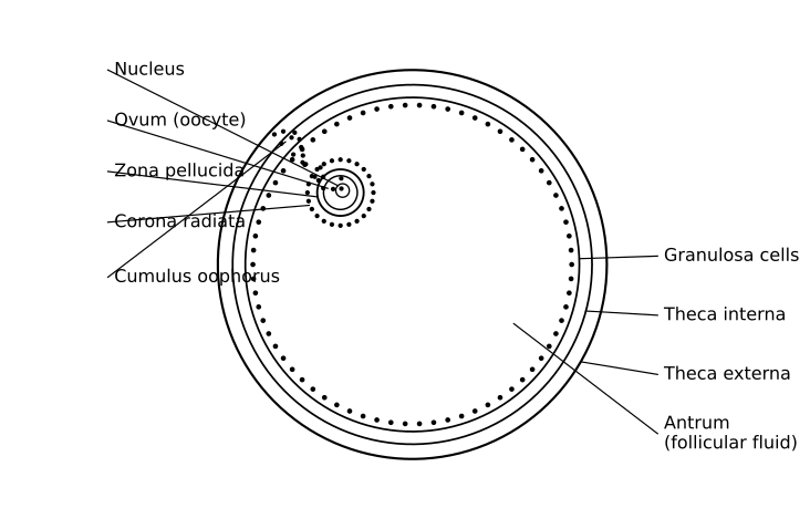
Draw a neat, labelled diagram of a mature Graafian follicle. Name the hormone responsible for triggering ovulation and state the source of this hormone.

Diagram for question 16: Human Reproduction
Show answer
Diagram of a mature Graafian follicle: (1 mark)

```
┌─────────────────────────────────┐
│ GRAAFIAN FOLLICLE │
│ │
Theca externa ──▶│╔═══════════════════════════════╗│
Theca interna ──▶│║ ╔═══════════════════════════╗║│
Granulosa cells ──▶│║ ║ ● ● ● ● ● ● ● ● ● ● ● ║║│
│║ ║ ● ● ● ● ● ● ● ● ● ● ● ║║│
Antrum (fluid)──▶│║ ║ (Follicular ║║│
│║ ║ Fluid) ║║│
│║ ║ ╔══════════════╗ ║║│
Cumulus ─────────│║ ║ ║●●●●●●●●●●●●║ ║║│
oophorus ─│║ ║ ║ ╔══════╗ ║ ║║│
Primary oocyte──▶│║ ║ ║ ║Oocyte║ ║ ║║│
(arrested at MI) │║ ║ ║ ╚══════╝ ║ ║║│
Zona pellucida──▶│║ ║ ║●●●●●●●●●●●●║ ║║│
│║ ║ ╚══════════════╝ ║║│
│║ ╚═══════════════════════════╝║│
│╚═══════════════════════════════╝│
└─────────────────────────────────┘
```

Required labels (any 4 for full diagram credit):
• Theca externa (outermost fibrous layer)
• Theca interna (inner vascular layer)
• Granulosa cells (layer surrounding antrum)
• Antrum (fluid-filled cavity containing follicular fluid)
• Cumulus oophorus (mound of granulosa cells supporting oocyte)
• Primary oocyte (arrested in Prophase I / Metaphase I)
• Zona pellucida (glycoprotein coat around oocyte)

---

Hormone responsible for triggering ovulation: (1 mark)
• Luteinising Hormone (LH) — specifically a SHARP SURGE (LH surge) in LH levels around day 13–14 of the menstrual cycle triggers ovulation.

Source of this hormone: (1 mark)
• LH is secreted by the anterior pituitary gland (adenohypophysis).

[Total: 1 (diagram with labels) + 1 (hormone name) + 1 (source) = 3 marks]
Q17Short Answer3 marks

During pregnancy, a cascade of hormones is secreted to maintain the uterine environment and prepare the body for parturition and lactation. Answer the following:
(i) Name the hormone responsible for triggering parturition. State its source and explain why the process of parturition is described as a 'positive feedback' mechanism. (2 marks)
(ii) Name TWO hormones involved in lactation after parturition. For each, state its source and specific role. (1 mark)

Show answer
PART (i) — Parturition [2 marks]

Hormone responsible for triggering parturition: Oxytocin
Source: Posterior pituitary gland (neurohypophysis) of the mother.

Positive Feedback Mechanism of Parturition:
• Near the end of gestation (~9 months), the fully developed foetus sends neuro-endocrine signals to the maternal system.
• These signals stimulate mild uterine contractions (foetal ejection reflex).
• Mild contractions → stimulate release of oxytocin from the posterior pituitary.
• Oxytocin → causes stronger and more forceful uterine contractions.
• Stronger contractions → stimulate the release of even MORE oxytocin.
• This forms a POSITIVE FEEDBACK LOOP: the response (contraction) amplifies the original stimulus, leading to increasingly intense contractions until the baby is expelled.
• Process ends only after delivery of the baby and placenta — stimulus is removed.

[Award 1 mark for: Oxytocin + posterior pituitary + foetal ejection reflex/initial contractions; Award 1 mark for: correct positive feedback loop explanation — each contraction → more oxytocin → stronger contraction, self-amplifying until delivery]

────────────────────────────────────────
PART (ii) — Hormones in Lactation [1 mark]

┌──────────────────┬─────────────────────────────────┬──────────────────────────────────────────────────┐
│ Hormone │ Source │ Role in Lactation │
├──────────────────┼─────────────────────────────────┼──────────────────────────────────────────────────┤
│ Prolactin │ Anterior pituitary (adenohypo- │ Stimulates the mammary glands to PRODUCE │
│ │ physis) │ (synthesise) milk (milk production / galactopoiesis)│
├──────────────────┼─────────────────────────────────┼──────────────────────────────────────────────────┤
│ Oxytocin │ Posterior pituitary │ Stimulates EJECTION (let-down / release) of milk │
│ │ (neurohypophysis) │ from the mammary glands during suckling │
└──────────────────┴─────────────────────────────────┴──────────────────────────────────────────────────┘

[Award ½ mark for any ONE correctly named hormone with correct source; Award ½ mark for the other correctly named hormone with correct source and role. Both must be present for full 1 mark. Roles may be briefly stated.]

Note: Colostrum (milk secreted during first few days after delivery) is rich in antibodies (IgA) — mention earns bonus credit if examiner discretion allows, but is not required for full marks.
Q18Short Answer3 marks

A couple visits a fertility clinic. Medical tests reveal that the woman has normal ovarian function and produces healthy eggs, but her fallopian tubes are completely blocked due to a previous infection. Her husband has normal, motile sperms. The doctor explains that fertilisation cannot occur naturally in this case.

(i) Name the specific region in the fallopian tube where fertilisation normally occurs. (1 mark)
(ii) Which Assisted Reproductive Technology (ART) technique would be MOST suitable for this couple? Give ONE reason for your choice. (1 mark)
(iii) After successful fertilisation in the laboratory, at what developmental stage is the embryo typically transferred into the uterus, and why is this particular stage chosen for transfer? (1 mark)

Show answer
(i) Fertilisation normally occurs in the ampullary region (ampulla) of the fallopian tube (oviduct). [1 mark]

(ii) IVF-ET (In Vitro Fertilisation and Embryo Transfer) — commonly known as the 'test-tube baby' technique — is the most suitable ART.
Reason: Since both fallopian tubes are completely blocked, fertilisation cannot occur inside the body. In IVF-ET, fertilisation is carried out outside the body (in vitro) in the laboratory, bypassing the blocked tubes entirely. [1 mark]

(iii) The embryo is transferred into the uterus at the 8-celled stage (blastomere stage up to 8 cells) or, alternatively, at the blastocyst stage.
Reason: At the 8-celled stage, the embryo is at a suitable developmental stage to implant successfully into the uterine endometrium. Transferring at this stage (or blastocyst) ensures the embryo has undergone sufficient early cell divisions and is developmentally competent for implantation, maximising the chance of a successful pregnancy. [1 mark]

[Examiner note: Award 1 mark for blastocyst stage as an equally acceptable alternative for part (iii), as NCERT states embryos with more than 8 blastomeres are transferred into the uterus.]
Q19Short Answer3 marks

A 28-year-old woman undergoing infertility evaluation was told by her doctor that her fallopian tubes are completely blocked due to a past infection. The doctor explained that the egg is released from the ovary but cannot reach the uterus.

(i) Draw a neat labelled diagram of a Graafian follicle showing at least four labelled parts.
(ii) Name the process by which the egg is released from the Graafian follicle. Which hormone triggers this process?
(iii) Given the blocked fallopian tubes, explain why natural fertilisation is impossible in this woman and name ONE Assisted Reproductive Technology (ART) technique that can help her conceive.

Show answer
MARKING SCHEME (Total: 3 marks)

─────────────────────────────────────
(i) Labelled diagram of Graafian follicle [1 mark]
─────────────────────────────────────

Diagram (draw in answer book):

┌─────────────────────────┐
│ GRAAFIAN FOLLICLE │
└─────────────────────────┘

Theca externa ──────────────────────────────┐
Theca interna ─────────────────────────────┐│
Granulosa cells ──────────────────────────┐││
Antrum (fluid-filled cavity) ────────────┐│││
││││
┌───────────────────────────────────┐ ││││
│ Theca externa (outermost layer) │◄──┘│││
│ ┌───────────────────────────┐ │ │││
│ │ Theca interna │◄───┘ ││
│ │ ┌─────────────────────┐ │ ││
│ │ │ Granulosa cells │◄─┘ │
│ │ │ ┌───────────────┐ │ │
│ │ │ │ ANTRUM │◄─┘ │
│ │ │ │ (follicular │ │
│ │ │ │ fluid) │ │
│ │ │ └───────────────┘ │
│ │ │ ↑ │
│ │ │ Cumulus oophorus │
│ │ │ (granulosa cells │
│ │ │ around oocyte) │
│ │ │ ↑ │
│ │ │ Secondary oocyte (n) │
│ │ │ + Zona pellucida │
│ │ └─────────────────────┘ │
│ └───────────────────────────────────┘ │
└───────────────────────────────────────┘

Required labels (any FOUR for full mark):
• Theca externa
• Theca interna
• Granulosa cells
• Antrum (follicular fluid)
• Secondary oocyte / primary oocyte
• Zona pellucida
• Cumulus oophorus

[Award 1 mark for correctly drawn and labelled diagram with minimum 4 labels]

─────────────────────────────────────
(ii) Process and hormone [1 mark]
─────────────────────────────────────

• Process: Ovulation [½ mark]
• Hormone that triggers ovulation: LH (Luteinising Hormone) — specifically the LH surge (sharp rise in LH level around day 14 of the menstrual cycle) [½ mark]

[Award 1 mark for both correct: process name + hormone name]

─────────────────────────────────────
(iii) Reason fertilisation is impossible + ART technique [1 mark]
─────────────────────────────────────

Reason:
• Fertilisation normally occurs in the ampullary region (ampulla) of the fallopian tube (oviduct).
• Since both fallopian tubes are blocked, the released secondary oocyte (egg) cannot travel toward the uterus, AND the sperms cannot reach the egg.
• Therefore, the sperm and egg cannot meet → natural fertilisation is impossible. [½ mark]

ART technique:
• IVF-ET (In Vitro Fertilisation – Embryo Transfer) / Test tube baby technique
— Egg collected from ovary and fertilised with sperm outside the body (in vitro) → embryo transferred to uterus, bypassing the blocked tubes. [½ mark]

[Award 1 mark for correct reason AND correct ART technique with brief explanation]

─────────────────────────────────────
VALUE POINTS SUMMARY:
(i) Diagram with ≥4 correct labels = 1 mark
(ii) Ovulation (½) + LH surge/LH (½) = 1 mark
(iii) Fertilisation site blocked/sperm-egg cannot meet (½) + IVF-ET / any correct ART with indication (½) = 1 mark
TOTAL = 3 marks
─────────────────────────────────────
Q20Short Answer3 marks

A 28-year-old woman underwent a laparoscopic procedure during which both her fallopian tubes were found to be completely blocked due to a previous infection. Her ovaries and uterus were reported to be fully functional by the surgeon.
(i) Name the specific region of the fallopian tube where fertilisation normally occurs. Why would a complete tubal blockage prevent fertilisation? (1 mark)
(ii) Despite the blockage, the couple wishes to have a biological child. Name ONE Assisted Reproductive Technology (ART) technique most suitable for this couple. Give ONE reason why this technique is appropriate in her case. (1 mark)
(iii) After successful fertilisation using ART, the zygote undergoes cleavage. State the stage at which the embryo is typically transferred to the uterus for implantation, and name the uterine layer in which it embeds. (1 mark)

Show answer
Marking Scheme (3 × 1 mark)

(i) Site of normal fertilisation + reason for blockage: (1 mark)
• Fertilisation normally occurs in the ampullary region (ampulla) of the fallopian tube (isthmo-ampullary junction).
• A complete tubal blockage prevents the ovulated ovum (released from the ovary) and the capacitated spermatozoa (ascending from the uterus) from meeting at the ampulla — hence syngamy cannot take place.
[Award ½ + ½ OR full 1 mark if both the site name and the reason are correctly stated. Award ½ if only the site OR only the reason is given.]

(ii) Suitable ART technique + reason: (1 mark)
• Technique: IVF-ET (In Vitro Fertilisation and Embryo Transfer) — also called the 'test tube baby' technique.
• Reason: In IVF-ET, the ovum and sperm are allowed to fertilise OUTSIDE the body (in vitro), completely bypassing the blocked fallopian tubes; the embryo is then transferred to the functional uterus.
[Award ½ for correct ART name + ½ for appropriate reason. GIFT (Gamete Intra-Fallopian Transfer) is NOT appropriate here because both tubes are blocked — do NOT award marks if GIFT is chosen without qualification.]

(iii) Stage of embryo transfer + uterine layer: (1 mark)
• The embryo is transferred to the uterus at the blastocyst stage (32–64 cell stage, formed after morula → blastocyst).
(Note: CBSE also accepts 8-cell morula stage for ZIFT into fallopian tube; however, since both tubes are blocked, transfer is directly to the uterus, so blastocyst is the correct and expected answer here.)
• The blastocyst embeds (implants) in the endometrium (inner lining / stratum functionalis of the uterus).
[Award ½ for blastocyst + ½ for endometrium. Award 0 if 'myometrium' or 'perimetrium' is written.]

[Examiner's Note — Common errors to watch for:
• Writing 'isthmus' as the site of fertilisation — incorrect; isthmus is where the sperm are stored temporarily, NOT where fertilisation occurs.
• Choosing GIFT or ZIFT as the ART technique without acknowledging that BOTH tubes are blocked — such answers must not receive full credit.
• Writing 'implantation in myometrium' — penalise; the myometrium is the muscular layer, not the receptive layer for implantation.]
Q21Short Answer3 marks

A couple is trying to conceive. Medical investigation reveals that the female partner has a serum progesterone level that drops sharply on Day 22 of her menstrual cycle, well before the expected next menstruation. Give reasons for the following based on this clinical finding:
(i) Why would implantation most likely fail in this case?
(ii) Why would measuring progesterone levels be a useful indirect indicator of whether ovulation has occurred?
(iii) Name the structure whose inadequate functional activity is directly responsible for the low progesterone in this scenario.

Show answer
Answer (3 marks — 1 mark each for parts i, ii, iii):

(i) Implantation failure due to premature progesterone drop: [1 mark]
• After fertilisation, the blastocyst must implant into a highly vascularised, glandular endometrium (uterine wall).
• Progesterone (secreted by the corpus luteum) is essential for maintaining the endometrial lining in a secretory/receptive state — it promotes glandular secretions and prevents menstruation.
• If progesterone drops sharply on Day 22, the endometrium undergoes premature breakdown (akin to early menstruation), making it non-receptive.
• Therefore, even if fertilisation has occurred, the blastocyst will not find a suitable endometrial environment for implantation, and pregnancy will fail.

(ii) Progesterone as indirect indicator of ovulation: [1 mark]
• Ovulation (release of the secondary oocyte) is triggered by the LH surge (around Day 14).
• After ovulation, the ruptured Graafian follicle transforms into the corpus luteum (under the influence of LH).
• The corpus luteum secretes progesterone — so a rise in serum progesterone in the post-ovulatory (luteal) phase (Days 15–28) confirms that a corpus luteum has formed, which in turn confirms that ovulation has taken place.
• If progesterone remains low throughout the cycle, ovulation has most likely not occurred (anovulatory cycle).

(iii) Structure responsible for low progesterone: [1 mark]
• Corpus luteum (formed from the ruptured Graafian follicle after ovulation).
• Inadequate corpus luteum function (luteal phase defect) leads to insufficient progesterone secretion, causing premature endometrial breakdown.

[Examiner note: Accept 'corpus luteum' for part (iii). For parts (i) and (ii), award 1 mark if the student correctly links progesterone → endometrium maintenance (i) and corpus luteum formation post-ovulation → progesterone rise (ii). Deduct no marks for spelling of 'corpus luteum' if clearly identifiable.]
Q22Long Answer5 marks

With reference to human reproduction, answer the following:
(i) Draw a labelled diagram of a mature human embryo sac (female gametophyte) and name the cells that directly participate in double fertilisation. (2 marks)
(ii) Trace the fate of the two products formed after double fertilisation, stating the ploidy of each product. (2 marks)
(iii) Give one reason why the endosperm must develop before the embryo in a fertilised ovule. (1 mark)

Show answer
MARKING SCHEME — Q (LA, 5 marks)

─────────────────────────────────────
PART (i) — Labelled diagram of mature embryo sac + cells participating in double fertilisation [2 marks]
─────────────────────────────────────

DIAGRAM (1 mark for correct drawing + 1 mark for correct labelling of all required parts):

```
MICROPYLAR END (top)
┌─────────────────────┐
│ Filiform apparatus │
│ ┌───┐ ┌───┐ │
│ │ S │ │ S │ │ ← Synergids (2)
│ └───┘ └───┘ │
│ ┌─────┐ │
│ │ Egg │ │ ← Egg cell (1)
│ │cell │ │
│ └─────┘ │
│ │
│ ┌─────────────┐ │
│ │ Central cell│ │ ← Central cell containing
│ │ ○ ○ │ │ 2 Polar nuclei (PNs)
│ └─────────────┘ │
│ │
│ ┌───┐ ┌───┐ ┌───┐│
│ │ A │ │ A │ │ A ││ ← Antipodal cells (3)
│ └───┘ └───┘ └───┘│
└─────────────────────┘
CHALAZAL END (bottom)
```

Required labels (any 6 for full credit):
• Egg cell — at micropylar end
• Synergids (2) — flanking egg cell (egg apparatus)
• Central cell — large central cell
• Polar nuclei (2) — inside central cell
• Antipodal cells (3) — at chalazal end
• Micropyle — opening at top
• Nucellus — surrounding tissue (if drawn)

[1 mark: correct 7-celled, 8-nucleate structure drawn]
[1 mark: minimum 5 parts correctly labelled with lead lines]

Cells directly participating in double fertilisation:
• Egg cell (n) — participates in syngamy ½ mark
• Central cell / the two polar nuclei (n + n) — participates in triple fusion ½ mark
(Award full 1 mark if both are named; do NOT award if only one is named)

Note: The synergids guide the pollen tube but do NOT directly fuse — do not credit them as participants in fertilisation.

─────────────────────────────────────
PART (ii) — Fate and ploidy of the two products of double fertilisation [2 marks]
─────────────────────────────────────

Product 1 — Zygote:
• Formed by: syngamy — male gamete (n) + egg cell (n) → Zygote (2n) [½ mark]
• Fate: Zygote undergoes mitotic divisions → develops into the Embryo (2n) [½ mark]

Product 2 — Primary Endosperm Nucleus (PEN):
• Formed by: triple fusion — male gamete (n) + 2 polar nuclei (n + n) → Primary Endosperm Nucleus (3n) [½ mark]
• Fate: PEN divides repeatedly → develops into Endosperm (3n), which provides nutrition to the developing embryo [½ mark]

Summary table (acceptable as an alternative to prose):

| Product | Ploidy | Develops into |
|---------|--------|---------------|
| Zygote | 2n (diploid) | Embryo |
| Primary Endosperm Nucleus (PEN) | 3n (triploid) | Endosperm |

(1×2 = 2 marks)

─────────────────────────────────────
PART (iii) — Why endosperm must develop before the embryo [1 mark]
─────────────────────────────────────

Accepted answer (any ONE of the following):

• The endosperm (3n) is a nutritive tissue; it must be established first so that it can supply nutrients (carbohydrates, proteins, fats) to the rapidly dividing embryo during its early development — without a pre-formed endosperm, the embryo would lack a nutritional source and could not develop normally. [1 mark]

OR

• The developing embryo is heterotrophic and cannot photosynthesize; the endosperm acts as the sole food reserve at this stage, so it must be formed before embryo differentiation begins. [1 mark]

─────────────────────────────────────
MARK SUMMARY
─────────────────────────────────────
Part (i): Diagram + labelling = 1 mark; Naming cells for double fertilisation = 1 mark → 2 marks
Part (ii): Fate + ploidy of zygote = 1 mark; Fate + ploidy of PEN = 1 mark → 2 marks
Part (iii): Reason for endosperm-first development = 1 mark → 1 mark
TOTAL = 5 marks
Q23Long Answer5 marks

Study the following and answer the questions given below:
(i) Draw a neat, labelled diagram of a mature human embryo sac (female gametophyte). Name all the cells present in it and state the ploidy of each cell type.
(ii) Describe the process of double fertilisation. Mention the ploidy of all the structures formed as a result of double fertilisation.

Show answer
PART (i): Labelled Diagram of Mature Human Embryo Sac (Female Gametophyte) [2 marks]

Draw a neat labelled diagram showing the following structures clearly:

[DIAGRAM — see description below]

MICROPYLAR END (top):
• Two Synergid cells flanking the egg cell (together forming the 'egg apparatus')
• One Egg cell (female gamete)

MIDDLE REGION:
• One large Central Cell containing two Polar Nuclei

CHALAZAL END (bottom):
• Three Antipodal cells

Labels that must appear on the diagram:
1. Synergids (×2)
2. Egg cell
3. Central cell
4. Polar nuclei (×2, inside central cell)
5. Antipodal cells (×3)
6. Micropyle
7. Chalaza

The mature embryo sac is 7-celled and 8-nucleate.

Cells present and their ploidy:

• Synergid cells (2 in number) — Haploid (n)
• Egg cell (1 in number) — Haploid (n)
• Central cell (1 in number, containing 2 polar nuclei) — Diploid (2n) [each polar nucleus is haploid (n), but the central cell as a whole is considered 2n]
• Antipodal cells (3 in number) — Haploid (n)

(1 mark for correct labelled diagram with all key structures; 1 mark for correctly naming all cells and stating ploidy of each)

---

PART (ii): Double Fertilisation [3 marks]

Double fertilisation is a unique characteristic feature of angiosperms. It involves two separate fusion events, both carried out by the two male gametes delivered by the pollen tube.

Process:

Step 1 — Pollen tube entry:
After pollination, the pollen grain germinates on the stigma and the pollen tube grows through the style and enters the ovule through the micropyle. The pollen tube releases two male gametes (each haploid, n) into the embryo sac.

Step 2 — Syngamy (First fertilisation):
One male gamete (n) fuses with the egg cell (n). This fusion is called syngamy. It results in the formation of the diploid zygote (2n), which later develops into the embryo.

Step 3 — Triple fusion (Second fertilisation):
The second male gamete (n) fuses with the diploid central cell (which contains two polar nuclei, each n). This fusion is called triple fusion (because one male gamete + two polar nuclei = three nuclei fuse). It results in the formation of the triploid Primary Endosperm Nucleus (PEN) (3n), which later develops into the endosperm.

Since fertilisation occurs twice — once with the egg cell and once with the central cell — the entire phenomenon is called Double Fertilisation.

Ploidy of structures formed as a result of double fertilisation:

• Zygote — Diploid (2n) [formed by syngamy: male gamete (n) + egg cell (n)]
• Primary Endosperm Nucleus (PEN) / Endosperm — Triploid (3n) [formed by triple fusion: male gamete (n) + two polar nuclei (n + n)]

(1 mark for explaining syngamy and formation of zygote with ploidy; 1 mark for explaining triple fusion and formation of PEN/endosperm with ploidy; 1 mark for correctly defining/describing double fertilisation as a whole and its significance as a characteristic of angiosperms)
Q24Long Answer5 marks

Rajan is a 28-year-old man who visited a fertility clinic with his wife after one year of unsuccessful attempts at conception. After clinical evaluation, the andrologist reported that Rajan's semen analysis showed: (i) very low sperm count, (ii) majority of sperm with immotile flagella due to absence of dynein arms, and (iii) significantly reduced levels of testosterone and FSH. The andrologist also noted that Rajan had a history of undescended testes (cryptorchidism) during childhood, which was surgically corrected at age 7.

(a) Trace the complete pathway of spermatogenesis, starting from spermatogonia to mature spermatozoa, mentioning the ploidy (2n/n) at each stage. (3 marks)
(b) Explain why cryptorchidism leads to impaired spermatogenesis, even if surgically corrected later. (1 mark)
(c) Based on the hormone profile given (low FSH and testosterone), identify which endocrine gland and which specific cell types are likely to be dysfunctional. Justify your answer. (1 mark)

Show answer
PART (a): Complete pathway of spermatogenesis with ploidy [3 marks]

Spermatogenesis occurs in the seminiferous tubules of the testes. The complete pathway is as follows:

1. Spermatogonia (2n) — Diploid germinal epithelial cells lining the seminiferous tubules. They undergo repeated mitotic divisions (multiplication phase) to increase in number. Some spermatogonia remain as stem cells; others differentiate into primary spermatocytes.

2. Primary Spermatocyte (2n) — A spermatogonium grows in size and undergoes DNA replication to become a primary spermatocyte. It then enters Meiosis I (reductional division).

3. Secondary Spermatocyte (n) — Formed after completion of Meiosis I. Each primary spermatocyte gives rise to two secondary spermatocytes. These are haploid (n) cells.

4. Spermatid (n) — Secondary spermatocytes undergo Meiosis II (equational division) to form four haploid spermatids from one primary spermatocyte. Spermatids are round, non-motile cells.

5. Spermatozoa (n) — Spermatids undergo spermiogenesis (morphological differentiation/metamorphosis) to form mature, motile spermatozoa. Excess cytoplasm is shed as residual bodies. Spermiation releases spermatozoa into the lumen of the seminiferous tubule.

Summary of ploidy at each stage:
• Spermatogonium → 2n
• Primary Spermatocyte → 2n
• Secondary Spermatocyte → n
• Spermatid → n
• Spermatozoon → n

[Value points: 1 mark for correct ploidy at each stage (2n for spermatogonium and primary spermatocyte; n for secondary spermatocyte, spermatid, spermatozoon) + 1 mark for correct sequence of stages + 1 mark for mentioning spermiogenesis/spermiation and the process of meiotic divisions leading to mature spermatozoa]

---

PART (b): Why cryptorchidism leads to impaired spermatogenesis even after surgical correction [1 mark]

Spermatogenesis requires a temperature approximately 2–2.5°C lower than the normal body temperature (37°C), which is why the testes are located in the scrotal sac outside the abdominal cavity. In cryptorchidism, the testes remain inside the abdominal cavity where the temperature is higher (~37°C). Prolonged exposure to this elevated temperature during the critical developmental period causes irreversible damage to the germinal epithelium (spermatogonia and Sertoli cells) of the seminiferous tubules. Even after surgical correction (orchiopexy) at age 7, the damage already sustained during the years of undescended testes cannot be fully reversed, resulting in permanently impaired spermatogenesis.

---

PART (c): Dysfunctional endocrine gland and cell types based on hormone profile [1 mark]

The dysfunctional endocrine gland is the anterior pituitary (adenohypophysis), and the dysfunctional cell types within the testes are the Sertoli cells and the Leydig cells (interstitial cells of Leydig).

Justification: FSH (Follicle Stimulating Hormone) is secreted by the gonadotroph cells of the anterior pituitary and normally acts on Sertoli cells of the seminiferous tubules to support spermatogenesis. Low FSH indicates reduced secretion from the anterior pituitary, leading to inadequate stimulation of Sertoli cells and consequently impaired sperm production. Additionally, LH (also from the anterior pituitary) normally stimulates the Leydig cells (interstitial cells) to secrete testosterone. Since FSH is low, it is likely that LH is also reduced (reflecting overall anterior pituitary gonadotroph dysfunction), resulting in insufficient stimulation of Leydig cells and hence significantly reduced testosterone levels. Thus, both Sertoli cells (responsible for nourishing developing spermatocytes) and Leydig cells (responsible for testosterone secretion) are dysfunctional due to inadequate gonadotropin stimulation from the anterior pituitary.

Want unlimited practice on Human Reproduction?

The full ClearSteps bank has 24+ reviewed questions on this chapter alone — with step-wise solutions, difficulty levels, and progress tracking. Verify your WhatsApp number and your free trial starts right here.

CBSE · CLASS 12
🎟️14-day free trial · code PRAC auto-applied
Mobile Number
+91
OTP will be sent to your WhatsApp

⭐ Trusted by 12,000+ students across India

Human Reproduction — Class 12 Biology Practice Questions